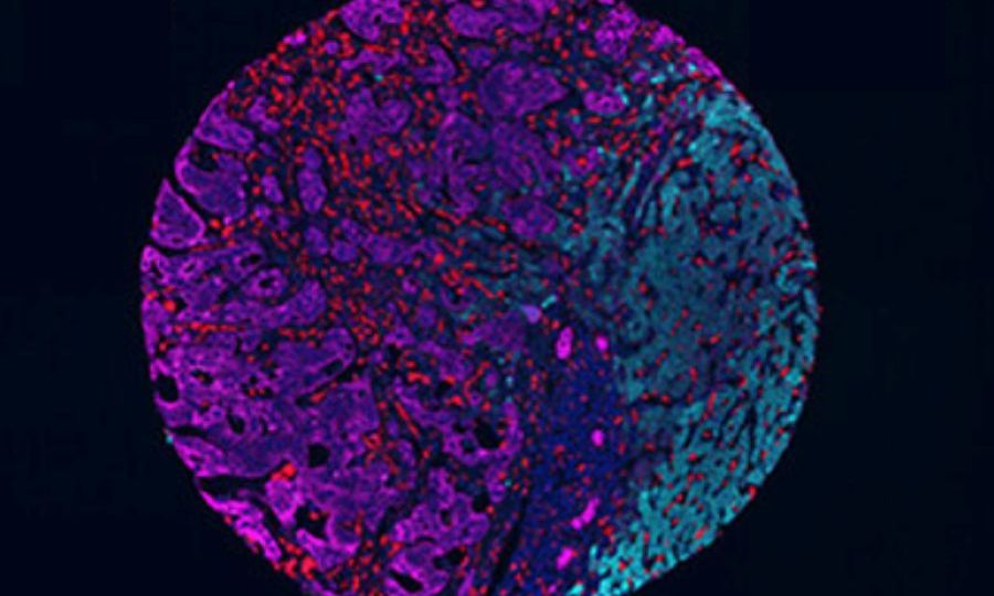
A recent study has provided new insights into how the tumour microenvironment may affect the development of liver metastases in patients with colorectal cancer. These metastases, which are one of the most serious complications of colorectal cancer, have different histological growth patterns. The research team led by Dr. David G. Molleví, head of the Tumour and Stromal Chemoresistance research group at IDIBELL and ICO, in collaboration with Dr. Artur Mezheyeuski, researcher of the VHIO’s molecular Oncology group and of the University of Uppsala and VHIO, has found that patients with metastases that follow the encapsulating growth pattern (which have a fibrous capsule around, and represent 20% of cases) have a better prognosis than those patients with non-encapsulating metastases (without fibrous capsule, which account for 80% of cases), due to the different cellular composition of the microenvironment – meaning all the cells that accompany the tumour cells – which favours a better response of the immune system in the former group of patients.
The study, which also involved pathologists and surgeons from Bellvitge University Hospital, as well as medical oncologists, included more than 130 patients. In tumour samples from these patients, the differences in the composition of the tumour microenvironment between the two types of metastasis were analysed using an advanced technique that allows the simultaneous detection of different proteins in the same tissue. Cytotoxic T lymphocytes, other immune system cells such as macrophages and also cancer-associated fibroblasts, which play an important role in disease progression, were studied.
The results revealed that encapsulated metastases had a higher presence of tumour-fighting immune cells, such as cytotoxic T-lymphocytes, and type I collagen, which supports the immune response. However, unencapsulated metastases had more cells capable of promoting immunosuppression and tumour growth, such as some specific immune cell types and tumour-promoting fibroblast subtypes.
Ultimately, this study demonstrates that liver metastases from colorectal cancer have different compositions of the tumour microenvironment depending on the histological growth pattern: in essence, encapsulated metastases have fewer immunosuppressive cells and demonstrate better immune capacity, while those without an envelope have a more unfavourable tumour composition.
These results are highly relevant for optimising the design of personalised therapies for colorectal cancer patients with different types of metastases. Until now, given that almost 80% of cases developed non-encapsulating metastases with little immune activity, liver metastases had been considered ‘cold tumours’ or ‘immune deserts’. However, this study has corroborated that the remaining 20% of cases with encapsulating metastases have anti-tumour immunity that could be exploited by immunotherapies. Therefore, predicting the type of histological growth of metastases could have a major impact on the personalisation of treatments. In this sense, the research group itself, in collaboration with researchers from VHIO’s Radiomics group and Belgian researchers, is working to predict the metastatic type using imaging techniques.
Reference


