- Researchers from the VHIO, promoted by the “la Caixa” Foundation, have verified in patients with advanced tumours that a new non-invasive tool, based on radiomics and the use of artificial intelligence, can help to predict the response to immunotherapy treatment with an accuracy of up to 75%.
- According to a study published in the journal Radiology, this new model can help to identify those patients who are most likely to benefit from immunotherapy.
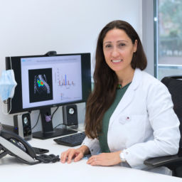
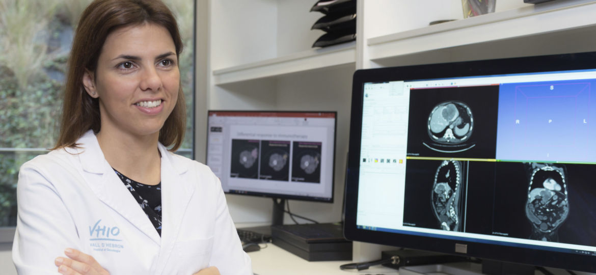
Barcelona, January 26, 2021- One of the current challenges in oncology is to best identify which cancer patients will respond to immunotherapy. The markers that exist to date are imperfect and have variable results depending on the type of tumour. A study, promoted by the “la Caixa” Foundation, is led by Dr Raquel Pérez-López, Principal Investigator of the Radiomics Group of the Vall d’Hebron Institute of Oncology (VHIO), which is part of the Vall d’Hebron Campus, has just been published in the journal Radiology. The study develops and validates a new radiological model based on the application of artificial intelligence models on pre-treatment computed tomography (CT) images.
In the results of its research, the team, led by Dr Raquel Pérez-López, has been able to verify how, in patients with advanced solid tumours, this tool was able to predict the response to treatments with anti-PD-1 and PD-L1 immunotherapeutic drugs with great sensitivity. “We were able to see that, in patients with bladder cancer, this sensitivity was 85% and in those with lung cancer it was 76%. The moderately high sensitivity of this test indicates a potential to better identify patients who may benefit from immunotherapy and, therefore, in whom this treatment may have priority over others,” explains the VHIO researcher.
This study was carried out at the Molecular Cancer Therapy Research Unit (UITM) – “la Caixa” Foundation, an international reference centre in the development of new cancer treatments and in the improvement of existing therapies, as well as in the optimal molecular selection of patients likely to respond to these therapies, through the development of advanced molecular diagnostic panels.
The newly developed tool is based on the analysis of tumour images obtained by means of a computed tomography (CT) scan prior to starting treatment. Through artificial intelligence models, associations can be established between the image and molecular profiles related to the immune response. “The amount of information that can be extracted by artificial intelligence from CT images is infinitely greater than that which can be extracted only with the observation of an expert. In this way, we have obtained a predictive score of the efficacy of immunotherapy in patients,” comments Dr Raquel Pérez-López, and continues: “In addition, this non-invasive tool will allow a more appropriate follow-up of the tumour’s progression and treatment over time and, furthermore, it allows us to evaluate the tumour in its entirety, not only at the biopsy sites.”
In reality, the future of precision medicine involves the combination of all the information obtained from multimicrobial platforms–where different analyses are combined, such as genes (genomics), proteins (proteomics), metabolites (metabolomics) or medical radiographic images (radiomics) among others–, to obtain a photo that is as personalised as possible, and conducive to the best decision-making and adjustment to the treatment, so as to offer the greatest benefit to patients at each stage of progression of their tumour.
A little explored path
The development of immunotherapy has been one of the great advances in the treatment of cancer in recent years. Specifically, the emergence of immune checkpoint inhibitors has been one of its great successes. These are a type of medicine that blocks proteins called checkpoints, which are made by some types of immune system cells, such as T cells, and certain cancer cells. These checkpoints help to prevent immune responses from being very strong. When their action is blocked, the T cells of the immune system destroy cancer cells better.
However, not all patients respond in the same way to these drugs and severe adverse effects can occur in some. Although work is being carried out on the development of new agents to combine with these inhibitors and make tumours more sensitive to these drugs, the development of markers that allow the optimal identification of tumours more sensitive to these treatments and that stratify patients appropriately to maximise the benefits of these therapies, minimise their risks, and accelerate the development of new drugs, is still much needed.
To date, various markers have been used for this identification, such as the expression of PD-L1 in the tumour, the instability of the microsatellites or the mutational load of the tumour, although the results of all of them are imperfect. Advances in personalised medicine are based on molecular characterisation based on genomics and proteomics. However, in order to perform these analyses, tissue samples need to be taken through biopsies or invasive surgeries. This represents a new limitation, in addition to not offering a general view of the tumour, which may be another obstacle considering its heterogeneity.
“Radiomics offers us a new way of analysing patients that overcomes these difficulties, as it is a non-invasive tool that offers us a complete view of the tumour, and also makes it possible to monitor the disease progression. The analysis of the images has enormous potential that has not yet been fully explored,” concludes Dr Raquel Pérez-López, who goes on to say that, despite the promising results of the research that are now being published, more studies are still needed in specific tumour types, and as new immunotherapies emerge. In this regard, Dr Raquel Pérez-López has been selected by the CRIS Research Talent Programme of CRIS against cancer, to lead one of the two programmes that have been convened this year. The programme will complete these studies. Specifically, this project will focus on seeking how to improve current imaging techniques, combined with genomics, which are used both in the diagnosis and monitoring of cancer.
In summary, Dr Raquel Pérez-López explains: “Through computational analysis, the images are processed obtaining data about the tumour that are impossible to perceive and analyse via the naked eye. In this way, we can integrate all this information hidden in the images into multiomic models while striving to improve our knowledge of cancer and the treatment of our patients. This will be the future,” she concludes.
Reference:
Marta Ligero, Alfonso Garcia-Ruiz, Cristina Viaplana, Guillermo Villacampa, Maria V. Raciti, Jaid Landa, Ignacio Matos, Juan Martin-Liberal, Maria Ochoa-de-Olza, Cinta Hierro, Joaquin Mateo, Macarena Gonzalez, Rafael Morales-Barrera, Cristina Suarez, Jordi Rodon, Elena Elez, Irene Braña, Eva Muñoz-Couselo, Ana Oaknin, Roberta Fasani, Paolo Nuciforo, Debora Gil, Carlota Rubio-Perez, Joan Seoane, Enriqueta Felip, Manuel Escobar, Josep Tabernero, Joan Carles, Rodrigo Dienstmann, Elena Garralda, Raquel Perez-Lopez. A CT-based radiomics signature is associated with response to immune checkpoint inhibitors in advanced solid tumors. Radiology 2021; 00:1-11. https://doi.org/10.1148/radiol.2021200928
